Back Of Skull And Neck Anatomy - Human Head Skull And Cervical Vertebrae Rear View Stock Photo Download Image Now Istock - The skull provides attachments for numerous muscles.
Back Of Skull And Neck Anatomy - Human Head Skull And Cervical Vertebrae Rear View Stock Photo Download Image Now Istock - The skull provides attachments for numerous muscles.. The foramen magnum, housing the brainstem, is also a part of the occipital bone. The skull or known as the cranium in the medical world is a bone structure of the head. Muscle head anatomy vocal organ diagram female neck anatomy neck wireframe head neck human anatomy head artery anatomy face pharynx vector neck degree head anatomy 3d. The skull provides attachments for numerous muscles. Excluding ear ossicles, it is made of 22 bones.
The skull is the bony skeleton of the head. Consequently, this is the information and knowledge that is important for. The splenius muscles originate at the midline and run laterally and superiorly to their insertions. Top head neck anatomy flashcards ranked by quality. From supporting the head to containing the spinal cord and nerves as they emerge from the skull this structure does it all.
Muscles Of The Head Neck Anatomy Model Youtube from i.ytimg.com It contains an external occipital protuberance that can be felt on the back of your head. The physicians originally studying human anatomy thought the skull looked like an helmet. Appreciate the link neck and vertebral column; An area called the occiput. Clinically, surface anatomy is used to split the neck into anterior and posterior triangles which provide clues as to the location of specific structures. Anatomy of the head and neck. In the neck, the platysma when contracted throws the skin into oblique ridges parallel with the fasciculi of the muscle. The neck is the area between the skull base and the clavicles.
It attaches to the clavicle and scapula.
In radiology, the 'head and neck' refers to all the anatomical structures in this region excluding the central nervous system, that is, the brain and spinal co. The trapezius originates from the skull and spine of the upper back and neck. The anatomy of the neck. In this chapter, you will learn the anatomic basis for the clinical practice of dental assisting. It contains an external occipital protuberance that can be felt on the back of your head. From supporting the head to containing the spinal cord and nerves as they emerge from the skull this structure does it all. Anatomy for sculptors uses a different approach to information, making it visual and understandable. This article will help you understand key anatomical structures in the skull and spine, with the goal of helping you better understand your condition. The skull provides attachments for numerous muscles. It supports and protects the face and the brain. The skull is the bony skeleton of the head. In the neck, the platysma when contracted throws the skin into oblique ridges parallel with the fasciculi of the muscle. Demonstrate practical lab skills in anatomy and an appreciation of the ethics lecture:
Consequently, this is the information and knowledge that is important for. Neck anatomy explained the neck begins at the base of the skull and connects to the thoracic spine the upper back. Anatomy of the head and neck. Excluding ear ossicles, it is made of 22 bones. An area called the occiput.
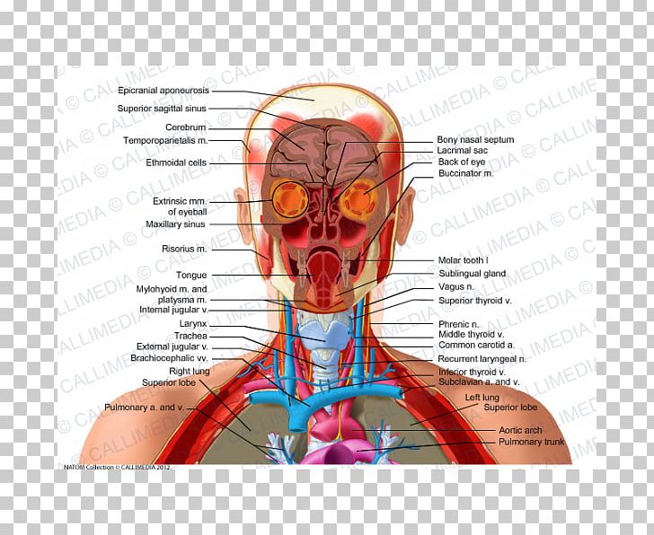
Neck Anatomy Pictures Bones Muscles Nerves from www.healthpages.org Anatomy of the head and neck. From the sides and the back of the neck, the splenius capitis inserts onto the head region, and the splenius. The upper part of your neck and the back of the head contain occipital nerves that can become very painful when irritated. Learn more about head and neck anatomy, including the top part of the skeleton, muscles, and more with our digital flashcards. From supporting the head to containing the spinal cord and nerves as they emerge from the skull this structure does it all. Knowledge of the anatomy of the vasculature of the head and neck from the thorax to the skull base is critical to the approach to diagnosis and treatment of cerebrovascular disease. Head, neck, and back anatomy. The skull provides attachments for numerous muscles.
From the sides and the back of the neck, the splenius capitis inserts onto the head region, and the splenius.
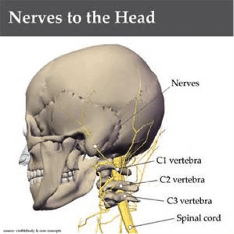
An area called the occiput. Apply anatomical knowledge in evaluating movement of the axial skeleton; The head rests on the top part of the vertebral column, with the skull joining at c1. Recurring pain in the back of your head at the base of your skull could be caused by occipital neuralgia. Anatomy, head neck anatomy, medical & nursing. Anatomy of the head and neck. 3 skull continued **fontanels in the skull are the unossified remnants of the membranes in newborns. Injury during delivery may also result in torticollis. The trapezius originates from the skull and spine of the upper back and neck. In the neck, the platysma when contracted throws the skin into oblique ridges parallel with the fasciculi of the muscle. From the sides and the back of the neck, the splenius capitis inserts onto the head region, and the splenius. Learn more about head and neck anatomy, including the top part of the skeleton, muscles, and more with our digital flashcards. While the bony framework of the neck is defined by the cervical.
How many moveable vertebrae are in the… what are the main purpose of transverse… In fact, there are twenty three in total, some of which are paired I will cover the basic forms of the mouth, some anatomical information, and the key information about the minor planes. Excluding ear ossicles, it is made of 22 bones. See more ideas about anatomy, skull anatomy, skull reference.
Back Of The Neck Anatomy Anatomy Drawing Diagram from cdn.imgbin.com The splenius muscles originate at the midline and run laterally and superiorly to their insertions. Top head neck anatomy flashcards ranked by quality. Injury during delivery may also result in torticollis. Clinically, surface anatomy is used to split the neck into anterior and posterior triangles which provide clues as to the location of specific structures. Recurring pain in the back of your head at the base of your skull could be caused by occipital neuralgia. I will cover the basic forms of the mouth, some anatomical information, and the key information about the minor planes. It supports and protects the face and the brain. We created the ★ ultimate anatomy study guide ★ to help you kick some gluteus maximus in any topic.
It attaches to the clavicle and scapula.
It contains an external occipital protuberance that can be felt on the back of your head. This article will help you understand key anatomical structures in the skull and spine, with the goal of helping you better understand your condition. The trapezius originates from the skull and spine of the upper back and neck. The head rests on the top part of the vertebral column, with the skull joining at c1. While the bony framework of the neck is defined by the cervical. All the bones of skull, joined together by sutures… anatomy ▶ head and neck ▶ bones and cartilages ▶ skull. In radiology, the 'head and neck' refers to all the anatomical structures in this region excluding the central nervous system, that is, the brain and spinal co. Appreciate the link neck and vertebral column; Despite being a relatively small region, it contains a range of important anatomical features. The muscles of the back and neck are responsible for maintaining posture and facilitating movement of the head and neck. This article describes the anatomy of the head and neck of the human body, including the brain, bones, muscles, blood vessels, nerves, glands, nose, mouth, teeth, tongue, and throat. Injury during delivery may also result in torticollis. How many moveable vertebrae are in the… what are the main purpose of transverse…
Learn more about head and neck anatomy, including the top part of the skeleton, muscles, and more with our digital flashcards back of skull anatomy. This anatomic region is complex and poses surgical challenges for otolaryngologists and neurosurgeons alike.


Post a Comment
0 Comments